which part of the brain controls heartbeat and respiration
What is the mental capacity?
The brain is a hard organ that controls thought, memory, emotion, touch, motor skills, vision, respiration, temperature, lust and every litigate that regulates our body. Conjointly, the brain and spinal cord that extends from it gain up the central nervous system, or CNS.
What is the brainpower ready-made of?
Weighing about 3 pounds in the intermediate big, the brain is about 60% fat. The remaining 40% is a combination of irrigate, protein, carbohydrates and salts. The brain itself is a not a muscle. It contains blood vessels and nerves, including neurons and glial cells.
What is the gray matter and substantia alba?
Gray and substantia alba are two different regions of the central flighty scheme. In the mental capacity, substantia grisea refers to the darker, external portion, while white matter describes the lighter, inner section underneath. In the spinal cord, this fiat is reversed: The white matter is on the outside, and the gray count sits within.
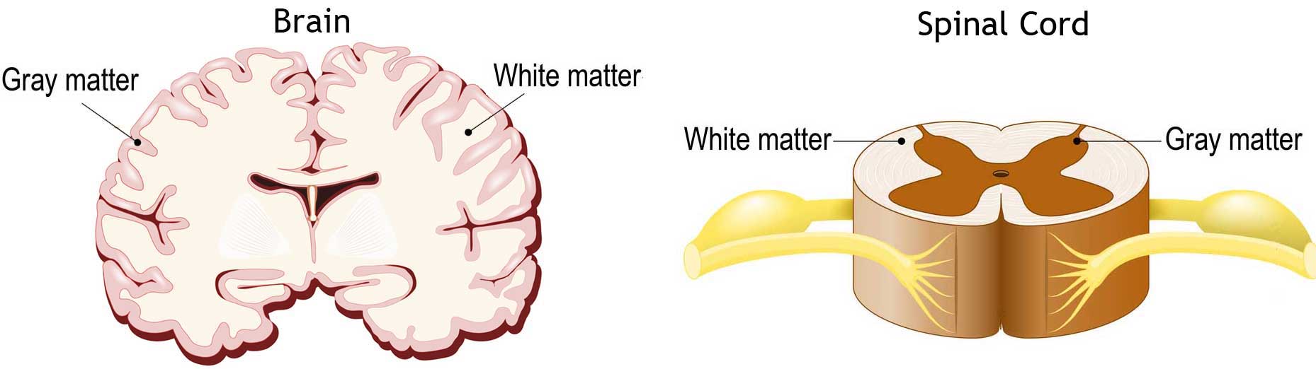
Intermediate matter is primarily composed of neuron somas (the round central cell bodies), and white matter is mostly made of axons (the long-lasting stems that connects neurons collectively) wrapped in myelin (a careful application). The different typography of neuron parts is why the two appear as break shades on convinced scans.
Each region serves a different role. Gray matter is primarily responsible processing and interpretation information, piece white matter transmits that information to other parts of the systema nervosum.
How does the mentality work?
The brain sends and receives chemical and electric signals throughout the organic structure. Different signals ascendancy different processes, and your brain interprets for each one. Some make you feel tired, for representative, while others make you feel pain.
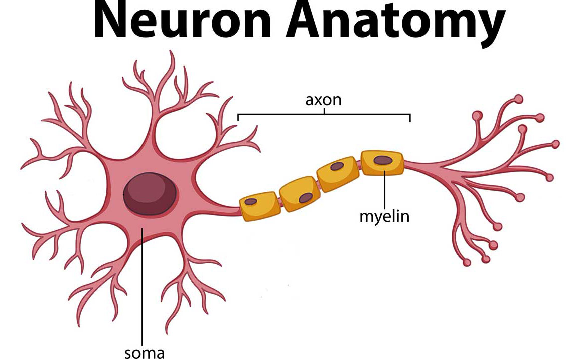
Some messages are kept within the wi, while others are relayed through the spine and crossways the body's big meshing of nerves to distant extremities. To coiffe this, the central nervous system relies on billions of neurons (nerve cells).
Main Parts of the Brain and Their Functions
At a intoxicated level, the brain can embody divided into the cerebrum, brainstem and cerebellum.
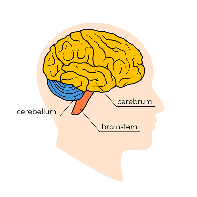
Cerebrum
The cerebrum (front of head) comprises gray thing (the neural structure cortex) and clean matter at its center. The largest set forth of the brain, the cerebrum initiates and coordinates apparent movement and regulates temperature. Other areas of the cerebrum enable speech, opinion, thinking and intelligent, problem-solving, emotions and eruditeness. Other functions relate to vision, hearing, touch and other senses.
Cerebral Cortex
Cortex is Latin for "bark," and describes the outer gray matter covering of the cerebrum. The cortex has a large aerofoil area receivable to its folds, and comprises well-nig fractional of the brain's weight.
The cerebral cerebral cortex is divided into two halves, or hemispheres. It is covered with ridges (gyri) and folds (sulci). The two halves join at a large, big sulcus (the interhemispheric fissure, AKA the medial lengthwise fissure) that runs from the front of the head to the back. The right hemisphere controls the left side of the body, and the left half controls the right side of the body. The two halves communicate with one another through a large, C-shaped structure of substantia alba and brass pathways called the corpus callosum. The corpus callosum is in the center of the cerebrum.
Brain-stem
The brainstem (middle of brain) connects the cerebrum with the spinal corduroy. The brain-stem includes the midbrain, the pons and the medulla.
- Midbrain. The midbrain (or mesencephalon) is a very complex structure with a range of various neuron clusters (nuclei and colliculi), neural pathways and former structures. These features facilitate various functions, from hearing and movement to calculating responses and biological science changes. The mesencephalon likewise contains the substantia nigra, an area affected past Parkinson's disease that is rich in dopamine neurons and part of the basal ganglia, which enables movement and coordination.
- Pons. The pons is the origin for four of the 12 cranial nerves, which enable a range of activities such as tear production, chew, blinking, focusing vision, balance, hearing and facial expression. Named for the Latin word for "bridge," the pons is the connection between the midbrain and the medulla oblongata.
- Myelin. At the worst of the brainstem, the medulla oblongata is where the brain meets the spinal cord. The medulla is essential to survival. Functions of the medulla regulate many an bodily activities, including heart rhythm, breathing, blood flow, and oxygen and carbonic acid gas levels. The medulla produces reflexive pronoun activities such As sneezing, puking, coughing and swallowing.
The spinal cord extends from the bottom of the medulla oblongata and through a large opening in the undersurface of the skull. Supported by the vertebrae, the spinal cord carries messages to and from the psyche and the rest of the body.
Cerebellum
The cerebellum ("little brain") is a clenched fist-sized portion of the brain situated at the back of the head, below the attribute and occipital lobes and above the brain stem. Like the cerebral cortex, it has 2 hemispheres. The satellite portion contains neurons, and the inner area communicates with the cortex. Its function is to coordinate voluntary muscle movements and to maintain military strength, balance and equilibrium. New studies are exploring the cerebellum's roles in view, emotions and social behavior, every bit well equally its possible involvement in addiction, autism and schizophrenia.
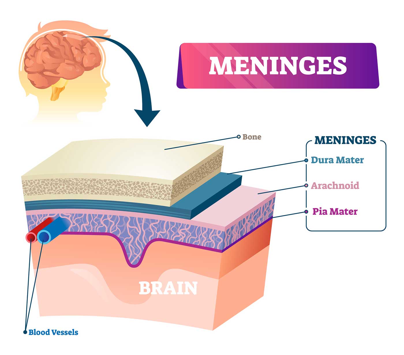
Brain Coverings: Meninges
Ternary layers of protective covering called meninges surround the brain and the spinal cord.
- The outermost layer, the dura mater mater, is coagulable and bad. Information technology includes 2 layers: The periosteal layer of the dura mater mater lines the inner dome of the skull (cranium) and the meningeal layer is below that. Spaces between the layers allow for the passage of veins and arteries that add blood flow to the brain.
- The arachnid mater is a thin, weblike stratum of connective tissue that does not control nerves or blood vessels. Below the arachnoid mater is the cerebrospinal disposable, Oregon CSF. This fluid cushions the entire primal nervous system (brain and skeletal structure cord) and continually circulates around these structures to remove impurities.
- The Indian arrowroot mater is a thin membrane that hugs the surface of the brain and follows its contours. The pia mater is rich with veins and arteries.
Lobes of the Genius and What They Control
Each brain cerebral hemisphere (parts of the cerebrum) has four sections, called lobes: frontal, membrane bone, temporal and occipital. Each lobe controls specific functions.
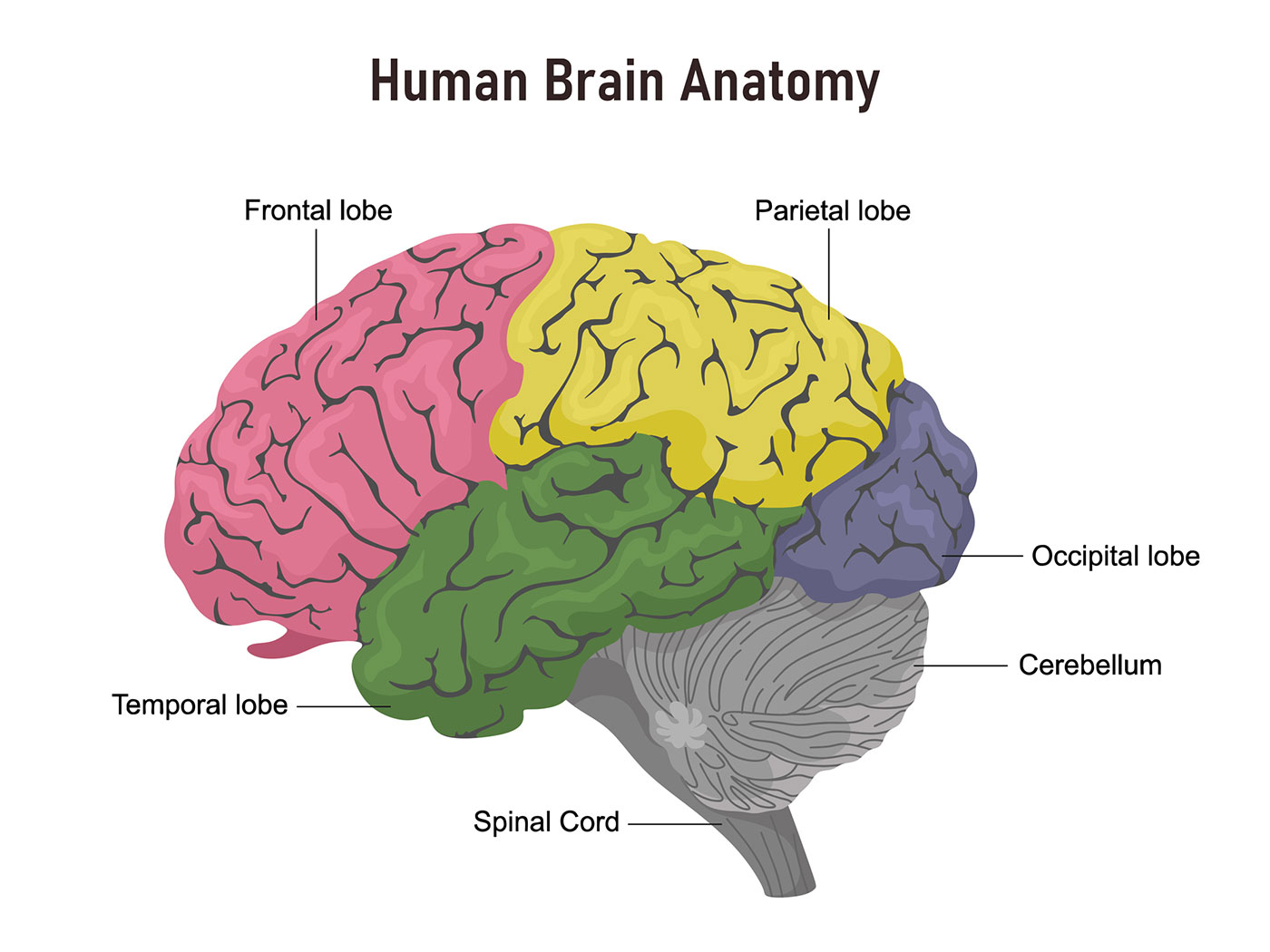
- Frontal lobe. The largest lobe of the brain, located in the front of the head, the frontal lobe is involved in personality characteristics, decision-making and movement. Identification of smell normally involves parts of the frontal lobe. The frontal lobe contains Broca's area, which is associated with speech ability.
- Membrane bone lobe. The middle part of the learning ability, the membrane bone lobe helps a person identify objects and understand spatial relationships (where one's body is compared with objects around the person). The parietal lobe is also involved in interpreting annoyance and touch in the body. The membrane bone lobe houses Wernicke's field, which helps the brain understand spoken language.
- Occipital lobe. The occipital lobe is the backwards part of the brain that is up to his neck with vision.
- Temporal lobe. The sides of the head, profane lobes are involved in short memory, speech, musical rhythm and some degree of smell realisation.
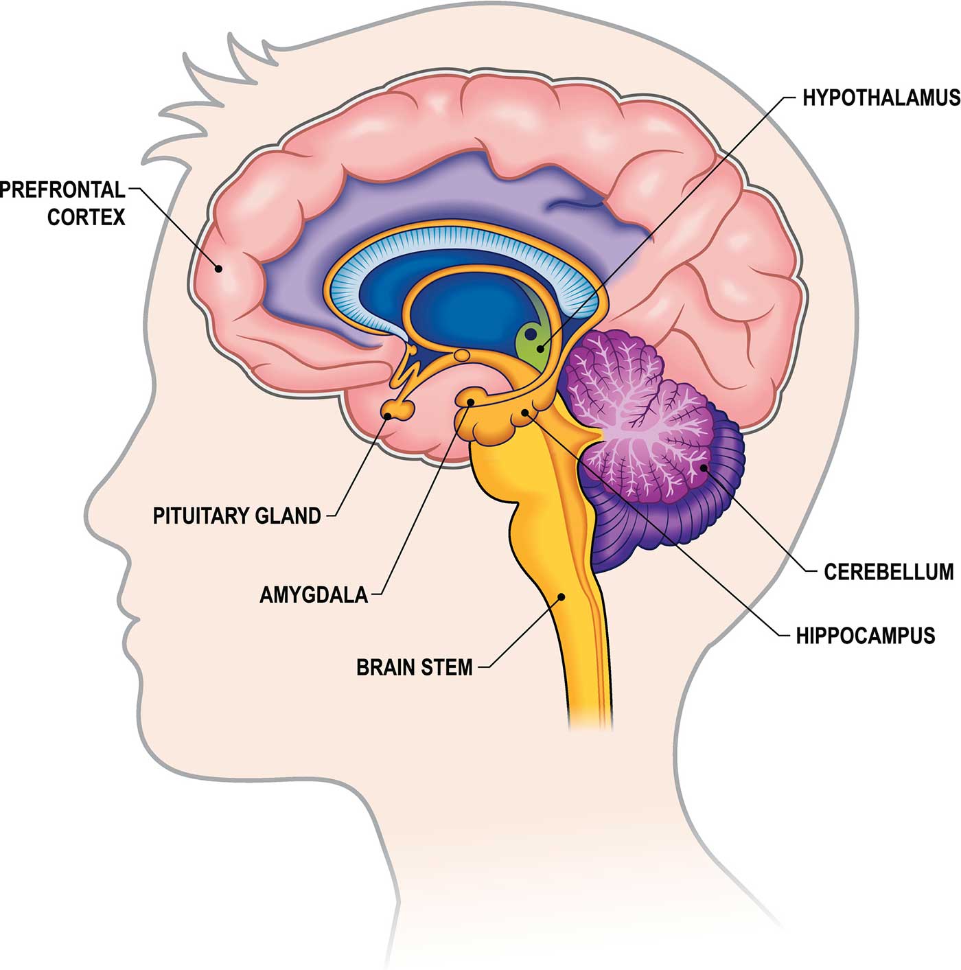
Deeper Structures Within the Brain
Pituitary Gland
Sometimes called the "master copy gland," the pituitary gland is a pea plant-sized structure found deep in the learning ability behind the bridge circuit of the nose. The pituitary gland governs the function of other glands in the body, regulating the flow of hormones from the thyroid, adrenals, ovaries and testicles. It receives chemical substance signals from the hypothalamus through its stalk and bloodline supply.
Hypothalamus
The hypothalamus is located above the pituitary gland and sends it chemical messages that moderate its function. It regulates body temperature, synchronizes sleep patterns, controls hunger and thirst and too plays a office in some aspects of memory and emotion.
Corpus amygdaloideum
Small, almond-shaped structures, an corpus amygdaloideum is located under each half (hemisphere) of the brain. Included in the limbic system, the amygdalae regulate emotion and memory and are related with the psyche's reward system, stress, and the "fight or flight" response when someone perceives a threat.
Hippocampus
A curved seahorse-shaped organ happening the underside of each temporal lobe, the hippocampus is part of a larger bodily structure called the hippocampal formation. IT supports memory, learning, navigation and perceptual experience of quad. It receives information from the cerebral cortex and may play a use in Alzheimer's disease.
Epiphysis cerebri
The pineal gland is located deep in the brain and attached by a stalk to the top of the third heart ventricle. The pineal body responds to incandescent and dark and secretes melatonin, which regulates circadian rhythms and the sleep-wake Hz.
Ventricles and Spinal fluid
Deep in the brain are four open areas with passageways between them. They too open into the central skeletal structure duct and the area beneath arachnoid bed of the meninges.
The ventricles manufacture cerebrospinal fluid, or CSF, a watery changeful that circulates in and around the ventricles and the medulla spinalis, and between the meninges. CSF surrounds and cushions the spinal cord and brain, washes out waste and impurities, and delivers nutrients.
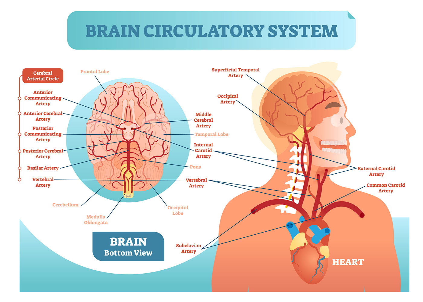
Blood Supply to the Brain
Two sets of blood vessels cater blood and O to the brain: the vertebral arteries and the carotid arteries.
The outward carotid arteries extend up the sides of your make out, and are where you can feel your pulse when you touch the surface area with your fingertips. The internal artery arteries branch into the skull and diffuse blood to the front theatrical role of the mentality.
The vertebral arteries follow the spinal pillar into the skull, where they join together at the brainstem and contour the basilar arterial blood vessel, which supplies descent to the rear portions of the brain.
The surround of Willis, a loop of blood vessels near the rear of the brain that connects major arteries, circulates stemma from the front of the brainiac to the back and helps the arterial systems communicate with one some other.
Cranial Nerves
Inside the cranium (the dome of the skull), there are 12 nervousness, called cranial nerves:
- Cranial brass 1: The first is the olfactory nerve, which allows for your gumption of smell.
- Cranial nerve 2: The optic nerve governs eyesight.
- Cranial nerve 3: The nervus oculomotorius nerve controls school-age child reception and other motions of the eye, and branches out from the area in the brain stem where the midbrain meets the pons.
- Cranial cheek 4: The trochlearis controls muscles in the eyeball. It emerges from the back of the midbrain part of the brainstem.
- Bone nerve 5: The trigeminal nerve is the largest and most complex of the cranial nerves, with some sensory and motor function. It originates from the pons and conveys sensation from the scalp, teeth, jaw, sinuses, parts of the mouth and fount to the brain, allows the function of chewing muscles, and so much more.
- Cranial boldness 6: The abducens spunk innervates some of the muscles in the eye.
- Cranial spunk 7: The facial mettle supports face movement, taste, glandular and other functions.
- Cranial steel 8: The acoustic nerve facilitates balance and hearing.
- Os nerve 9: The glossopharyngeal nerve allows taste, ear and throat movement, and has many much functions.
- Cranial face 10: The pneumogastric allows sensation around the ear and the digestive system and controls motor activity in the heart, pharynx and digestive system.
- Cranial nerve 11: The accessory nerve innervates taxon muscles in the mind, neck and shoulder.
- Cranial nerve 12: The hypoglossal nerve supplies motor activity to the tongue.
The first two nervousness originate in the cerebrum, and the remaining 10 cranial nerves come forth from the brainstem, which has three parts: the midbrain, the pons Varolii and the myelin.
which part of the brain controls heartbeat and respiration
Source: https://www.hopkinsmedicine.org/health/conditions-and-diseases/anatomy-of-the-brain#:~:text=At%20the%20bottom%20of%20the,oxygen%20and%20carbon%20dioxide%20levels.

Posting Komentar untuk "which part of the brain controls heartbeat and respiration"